The Washington University Musculoskeletal Research Center (MRC) and Center for Cellular Imaging (WUCCI) have partnered to acquire and operate a “3D X-ray microscope” for high-resolution imaging of a wide variety of biological samples (from bone to brain to kidney to fat) as well as synthetic biomaterials. This resource is now available to all members of the Washington University research community.
 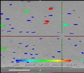 |   |  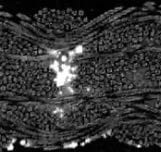 |
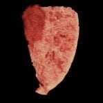
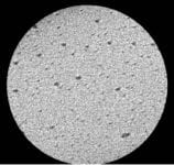
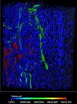
| Bone image (1.8 um resolution) reveals microvascular canals (red, green) and osteocyte lacunae (blue). (ZEISS, MRC & Silva lab) | Mouse embryo – multiple tissues stained with KI3 (James Fitzpatrick/WUCCI & Eghtesady Lab) | Woven biomaterial scaffold seeded with adult stem cells (bright). (Matt Joens/WUCCI & Guilak Lab) |
  |  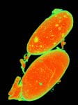 |
| Mouse Kidney – stained with Os / UA (Matt Joens/WUCCI & Humphreys Lab) | Mouse Kidney – stained with KI3 – showing cancer metasteses in green (Matt Joens/WUCCI & Weilbaecher Lab) |
The ZEISS Xradia 520 Versa 3D X-ray microscope (XRM) is capable of sub-micron resolution, high-contrast x-ray computed tomography (CT) imaging. The system has specifications on par with other “nanoCT” systems, and in addition has dual-energy and phase-contrast capabilities to extend the range of applications beyond radiodense materials. Its purchase was funded by the NIH Office of Research Infrastructure Programs through a High-End Instrumentation Grant (1 S10 OD021694-01) awarded to Dr. Matthew Silva, Department of Orthopaedic Surgery and Musculoskeletal Research Center. The system is operated the Department of Neurology and Center for Cellular Imaging, as part of a partnership between the MRC and WUCCI. It is located at the WUCCI (4515 McKinley Research Building, Washington University School of Medicine). For information, please contact the Simon Tang , WUCCI staff at wucci@wustl.edu, or visit wucci.wustl.edu.
Applications of 3D X-Ray Microscope
The XRM system is for ex vivo imaging of samples from < 1 mm to ~10 cm in size. There is a wide-range of sample geometries and imaging resolutions (voxel size) that are possible, and imaging settings (energy, filters, projections, integration time, etc.) are all under user control, allowing for great flexibility. The applications that we have already demonstrated with the XRM are shown below. This is an illustration of possible applications – we are excited to try new applications so please contact us if you have a project or idea.
1. Cortical Bone – porosity & microcracks
   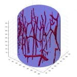 |  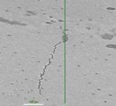 |
| (Left) 2D slice of cortical bone from rabbit ulna imaged at 0.99 um resolution reveals vascular and cellular (osteocyte) microporosities. (Right) 3D vascular porosity image from this sample. (Dan Leib, MRC & Silva Lab) | 2D slice of rat ulna imaged at 0.85um reveals microcrack after fatigue loading. (ZEISS, MRC & Silva Lab) |
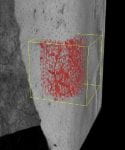  |   | 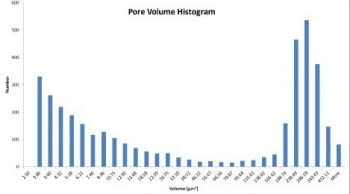  |
| XRM scan of mouse tibia imaged at 1.8 um reveals vascular and cellular microporosity. (Left) 3D view of cortex with porosity of sub-region shown in red. (Middle) Porosity color coded by volume – smaller osteocyte lacunae are blue; vascular pores are green or red. (Right) Histogram reveals distribution of porosity size. (ZEISS, MRC & Silva Lab) |
2. Bone marrow adipose tissue
 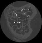 | 2D slice through mouse tibia imaged at 2.2 um reveals individual lipid droplets in the bone marrow (Osmium contrast; Dan Leib/MRC & Scheller/Craft Lab) |
3. Fracture Callus and Vascularity
3D reconstructions of mouse femur w/fracture callus. (Left) Mineralizedsample with low-density callus andvessels. (Right) Demineralized sampleshowing only vasculature. (Microfilcontrast; James Fitzpatrick/WUCCI,Dan Leib/MRC & Silva Lab)
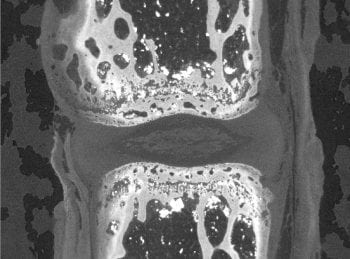
4. Whole Joints
 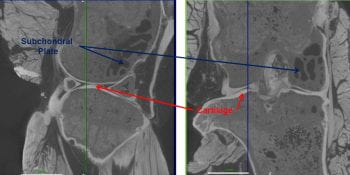 | Sagittal and coronal slices through mouse knee joint revealing mineralized and non-mineralized tissues, incl. subchondral bone (blue arrows), articular cartilage (red arrows), ligaments, joint capsule, meniscus and muscle. (Scanned at 4.5 um voxel size with PTA contrast; ZEISS, MRC & Silva Lab) |
  | Sagittal slice through rat elbow revealing humero-radial and humero-ulnar articulations. Anterior joint capsule is seen between humeral condyle and muscle. (Scanned at 4.9 um voxel size with PMA contrast; Dan Leib/MRC & Lake Lab) |
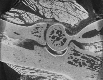
5. Vertebra & Intervertebral Disc
  | 2D slice of mouse tail vertebra-disc-vertebra reveals details of intervertebral disc (IVD) structure, including outer annulus, inner nucleus and endplates. (Scanned at 1.9 um voxel size with PMA contrast; Dan Leib/MRC & Tang Lab) |